Recorded Webinars

Presented By:
Tobias Gilk, MRSO, MRSE
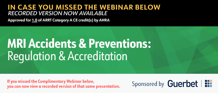
Despite the “safe imaging option” reputation, when it comes to MRI there are risks that need to be considered in the safety regulation and accreditation standards. Unlike ionizing radiology modalities, such as X-ray, CT, and fluoroscopy, MRI uses powerful magnetic fields and radio waves to produce detailed images of internal body tissues and structures. While MRI is generally considered safe, there are still risks associated with the use of this technology. To address these potential risks, safety regulations and accreditation standards for MRI must be carefully designed and implemented to respond to the safety concerns that are peculiar to MRI. Healthcare providers also need to have access to training and education on MRI accidents and safe practices.
Join Tobias Gilk, MRSO, MRSE for a deep dive into the critical topic of MRI safety regulation and accreditation standards.
This informative session will shed light on the gaps that exist in MRI safety regulation and accreditation, as compared to ionizing radiology. Participants will learn about the emergent risks associated with MRI and the preventative measures that can be taken. Additionally, Toby will also provide a scorecard of how well accreditation meets its promise of assuring safety when it comes to MRI. Participants will leave, equipped with valuable insights crucial to ensuring maximum safety in MRI practices.
Learning Objectives:
- Explain the long-term trajectory of MRI safety and MRI accidents.
- Identify a number of existing MRI safety best practices that are profoundly effective at preventing MRI injury.
- Quantify how many of the injury-preventing best practices are minimum required elements for hospital and MRI accreditation programs.
This activity may be available in multiple formats or from different sponsors. ARRT regulations state that an individual may not repeat an activity for credit if it was reported in the same or any subsequent biennium.

Presented By:
Ari D. Goldberg, MD, PhD
New MRI contrast agents continue to be developed, with a focus on continued improvements in stability and safety, as well as maximization of image clarity. These distinctions rest in the molecular structure of the particular gadolinium-based contrast agent. Understanding the relative safety and efficacy of these agents is important in the radiologic management of patients.
During this webinar, we’ll look closely at novel advances related to MRI contrast safety and management; understanding the risks associated with various agents as well as their efficacy.
Learning Objectives:
- Identify the different Gadolinium-based contrast agent classifications.
- Explain how Gadolinium-based contrast agent stability and safety is evaluated.
- Describe the concept of gadolinium-based contrast agent "relaxivity".
This activity may be available in multiple formats or from different sponsors. ARRT regulations state that an individual may not repeat an activity for credit if it was reported in the same or any subsequent biennium.

Presented By:
Jeffrey H Maki, MD, PhD
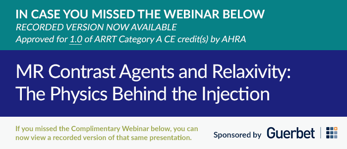
Gadolinium based contrast agents are vital to many clinical applications of MR imaging and contribute greatly to MRI’s powerful diagnostic abilities. As physicians who prescribe, or technologists who administer these contrast agents, it is important to have a good understanding of how they work, and what the differences are between various formulations or “brands”. Gadolinium is a highly toxic heavy metal and is only rendered safe for human administration by tightly binding it to a molecular structure called a chelate. Different gadolinium formulations are characterized by different chelates. The structure of this chelate dictates everything about the agent, from its biodistribution, to its safety profile to how well it does its job of increasing image contrast to highlight different forms of pathology.
Join Jeffrey H Maki, MD, PhD, for a webinar that will discuss the basics of MR relaxation times and how these impact image formation, the mechanism by which MR contrast agents work to relax protons, the different ways the structure of the chelate can be designed to improve this relaxivity and the potential of increased relaxivity contrast agents to impact clinical MR imaging.
At the end of this dynamic discussion, attendees will have a much better understanding of the changing landscape of gadolinium-based contrast agents and be better equipped to most effectively choose and use the most appropriate agent for their patient’s needs.
Learning Objectives:
After attending this webinar the participants will be better able to:
- Explain the relationship between MR relaxation times and contrast agent relaxivity.
- Describe what a chelate is and why it is important.
- Discuss two ways a chelate can be constructed to increase relaxivity.
- Rank the relaxivities and dosing of the currently available gadolinium contrast agents.
Approved for 1.0 of ARRT Category A CE credit(s) by AHRA
This CE activity may be available in multiple formats or from different CE sponsors. ARRT regulations state that an individual may not repeat a self-learning CE activity for credit if it was reported in the same CE biennium.

Presented By:
Donna Roberts, MD, MS
Magnetic resonance imaging (MRI) with gadolinium-based contrast agents (GBCAs) plays a critical role in the diagnosis, evaluation, and follow-up of many CNS disorders. However, as with all drugs, the risks versus benefits must be assessed prior to each use. GBCAs are widely used and have shown a very good safety profile. Although rare, acute hypersensitivity reactions, a condition known as nephrogenic systemic fibrosis in patients with abnormal renal function, and more recently the long-term retention of gadolinium in the body has been associated with GBCA use.
Join Donna Roberts, MD, MS, for a webinar that will discuss the current landscape of GBCA usage in patients with CNS disorders. In addition, the webinar will review the current practice guidelines and future directs for maximizing the clinical benefits of GBCAs while reducing the exposure of patients to GBCAs.
Learning Objectives:
After attending this webinar the participants will be better able to:
- Discuss clinical indications for the appropriate use of MRI contrast agents.
- Discuss considerations for selecting appropriate MRI contrast agents for specific patient populations including stability, efficacy, and patient factors.
- Describe the risks versus benefits associated with MRI contrast agent usage.
- Review recent updates to regulatory guidelines concerning MRI contrast agent usage.
Approved for 1.0 of ARRT Category A CE credit(s) by AHRA
This CE activity may be available in multiple formats or from different CE sponsors. ARRT regulations state that an individual may not repeat a self-learning CE activity for credit if it was reported in the same CE biennium.
Presented by:
 |
 |
| Peter McCaffrey, MD | Sina Aslan, PhD |
Healthcare institutions house vast amounts of imaging data that can fuel valuable collaborations with researchers and commercial entities; however, it takes considerable resources and lead time to create these data sets and share them in compliant and efficient ways. Dr. Peter McCaffrey at the University of Texas Medical Branch has implemented a platform for streamlining the creation of both radiology and pathology data sets, enabling more efficient collaborations both inside and outside the UT system. As a result, grant and publication timelines are accelerated, grant applications are more successful, and the health system’s data is being used in innovative new models.


PRESENTED BY:
Jacqueline K. Peccerillo, BS, R.T.(R)(CT)(BD)(PET)
Radiography Clinical Coordinator
Instructor of Radiography
Health care is an interdisciplinary field, and R.T.s are exposed to a wealth of alternative modalities and employment opportunities. Which ones should you choose and how do you move into a new role? By researching the current job market, R.T.s can analyze trending opportunities and profitable positions. After identifying opportunities, a self-assessment must be performed to determine goals, strengths, and weaknesses.
In this complimentary recorded webinar, Professor Jacqueline Peccerillo, R.T. (R)(CT)(BD)(PET), shares actionable strategies to uncover opportunities for professional growth and perform self-assessments to identify fitting opportunities. Professor Peccerillo also shares vignettes describing the journeys that other R.T.s have taken to reach their goals to help attendees take the next steps towards the wisest and most fitting career pathway enhancements.
Approved by the ASRT for Category A continuing education credit.
This CE activity may be available in multiple formats or from different CE sponsors. ARRT does not allow self-learning CE activities (e.g., Internet courses, home study programs, directed readings) to be repeated for CE credit in the same CE biennium.

PRESENTED BY:
Tobias Gilk, MRSO, MRSE
Apart from no-show patients, clearing patients with implants, medical devices, or foreign bodies is one of the most time-consuming and delay-producing aspects of MRI care. In this presentation Toby Gilk will walk through the various legal duties and limitations relative to authorizing patients with foreign bodies to proceed with an MRI exam, with examples of both appropriate and inappropriate guiding policies.
Next, Mr. Gilk will walk you through ‘untangling’ the risks of scanning patients with devices, reducing one unsolvable mess into identifiable separate risks associated with the three different electromagnetic fields of the MRI: Static Magnetic Field; Time-Varying Gradients; and Radiofrequency Magnetic Fields. Finally, he’ll provide you with a simplified risk-assessment tool that can help evaluate potential risks of harm for every possible implant, device, shrapnel or foreign body.
Approved by the ASRT for Category A continuing education credit.
This CE activity may be available in multiple formats or from different CE sponsors. ARRT does not allow self-learning CE activities (e.g., Internet courses, home study programs, directed readings) to be repeated for CE credit in the same CE biennium.
| Pediatric to Bariatric Imaging: Managing their unique complexities with Spectral-Detector CT |
|
PRESENTED BY:
 |
 |
| Dianna Bardo, MD University of Arizona School of Medicine |
Gopal Punjabi, MD Chair, Department of Radiology, Hennepin Healthcare Minneapolis MN |
Spectral-Detector CT has made a significant mark in imaging across all patient populations from Pediatric to Bariatric and has made unexpected and incidental findings, suboptimal contrast enhancement, blooming artifacts (calcified plaques, stents, etc.), Low eGFR and Metal implant complexities challenges of the past.
This is an Expert Forum Webinar with Dr. Dianna Bardo and Dr. Gopal Punjabi who share their experience utilizing Spectral-Detector CT and its clinical support tools to overcome the unique complexities and challenges that come when imaging pediatric and bariatric patients. They discuss their path in introducing this technology to their departments, and give examples of how it has helped them overcome imaging and diagnostic challenges with these unique patient types such as reducing contrast and radiation dose, eliminate repeat scans, subsequent imaging and follow up scans, optimize and standardize workflow, and enhance patient and staff experience. Click below to view the recorded version and learn how you can apply similar results to your own departments.
| View Recorded Version |
| The latest clinical trends in cardiac CT and MR, recent COVID-19 workflow implications and the power of Advanced Visualization and AI for related analysis |
|
PRESENTED BY:
 |
 |
 |
| Dr. Tim Leiner is Professor of Radiology and holds the Chair in Cardiovascular Imaging at Utrecht University Medical Center, Utrecht, The Netherlands. | Jonathon A. Leipsic, MD, FRCPC, MSCCT, is the Chairman of the Department of Radiology for Providence Health Care, Vancouver Coastal Health, Canada. | Kevin Lev is a Marketing Director for Advanced Visualization and AI Solutions at Philips. |
Recently, mounting clinical evidence confirming the accuracy, prognostic value, and role of CT and MR cardiac imaging in guiding clinical management has led to a shift in the guidelines. Now, cardiac CT and MR are recommended to provide first-line diagnostic insights in different clinical questions.
This webinar will elaborate on the latest clinical trends in cardiac CT (focusing on coronary artery disease) and MR, and how imaging workflows have been affected by the recent COVID-19 pandemic.
Finally, this session will touch upon the latest enhancements in automation, throughput and AI tooling in Advanced Visualization platforms supporting cardiac CT and MR analysis.
Webinar Learning Objectives
- Learn about the latest trends in cardiac CT & MR imaging
- Learn about the COVID-19 impact on cardiac CT & MR imaging
- Advanced Visualization offerings in the fields of cardiac CT & MR analysis
| View Recorded Version |


