 On the Case
By John Lausch; Rabita Alamgir, MD; Nikhil Kanthala, DO; and Laura Gregorio, MD
Radiology Today
Vol. 25 No. 8 P. 36
History
A 20-year-old woman presented with a 1.5-year history of vomiting and abdominal discomfort. Various treatments, including ondansetron, metoclopramide, and haloperidol, were ineffective, with only amitriptyline offering slight relief. Extensive testing, including a gastric emptying scan, upper endoscopy, hormone panels, and pelvic ultrasound, were already performed and were all unremarkable. Abdominal CT angiography (CTA) was then performed for further evaluation.
Findings
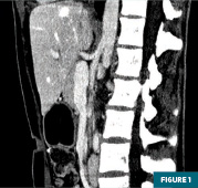
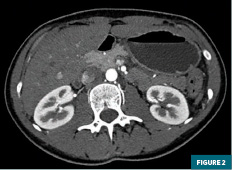
A sagittal abdominal CTA image (Figure 1) demonstrated a congenital celiacomesenteric trunk (CMT) with a common origin of the celiac trunk and the superior mesenteric artery (SMA), as well as a narrowed aortomesenteric angle (AMA) and distance of 17 degrees and 3 mm, respectively. An axial image (Figure 2) demonstrated dilatation of the stomach as well as the first and second portions of the duodenum, with relative lack of fat surrounding the SMA. There was significant caliber reduction and compression of the third portion of the duodenum between the SMA and the abdominal aorta. These findings are consistent with SMA syndrome.


Diagnosis
Congenital CMT with common origin of the celiac trunk and SMA associated with SMA syndrome
Discussion
SMA syndrome is a rare entity with an estimated incidence of 0.013% to 0.3%, characterized by the compression of the third portion of the duodenum between the SMA and the aorta.1,2 This compression can lead to symptoms such as nausea, vomiting, abdominal pain, and weight loss.3,4 SMA syndrome is more common in females and in those aged 10 to 39.5 Its diagnosis can be challenging due to its rarity and nonspecific presentation. In this case, the diagnostic process was further complicated by the presence of a congenital CMT, a rare vascular anomaly where the celiac trunk and the SMA share a common origin from the aorta. Our patient’s initial workup was unremarkable, although the abdominal CTA provided a conclusive evaluation by allowing for depiction of bowel and vascular anatomy.
The pathogenesis of SMA syndrome stems from an abnormal AMA and aortomesenteric distance (AMD). Studies have demonstrated the average AMA in the general population ranges between 28 and 65 degrees, while the average AMD ranges from 10 mm to 34 mm. Although these values can vary, one study has reported a sensitivity and specificity of 100% for an AMD <8 mm and 100% sensitivity with 42% specificity for an AMA <22 degrees in symptomatic patients.5 The fat surrounding the third portion of the duodenum, the typical bowel segment transversing between the SMA and aorta, is critical to maintaining this necessary angle and distance.1 SMA syndrome occurs when this angle and distance are reduced to the point of bowel obstruction. Reductions in AMA and AMD are caused by certain conditions and surgeries. Rapid weight loss from wasting diseases such as AIDS, malabsorption, cancer, and cachexia can diminish the protective cushion provided by the peritoneal fat around the duodenum.6 More commonly, corrective surgery for spine abnormalities such as scoliosis has been a well-documented cause of SMA syndrome secondary to spinal manipulation, causing traction and hyperextension of the spine, further reducing the AMA.7 Similarly, anatomical variations, such as an inferior origin of the SMA or ligament of Treitz malposition, can cranially displace the duodenum into the more acute proximal AMA.6
In our case, the patient’s CTA revealed an abnormal AMA of 17 degrees and an AMD of 3 mm. These findings, combined with her symptoms of postprandial vomiting and abdominal pain, supported the diagnosis of SMA syndrome. The presence of these radiographic and clinical findings in conjunction with the congenital CMT further complicated the clinical picture. While the CMT is typically an incidental finding, its coexistence with SMA syndrome, in this case, raises the question of whether the anomalous vascular anatomy may predispose patients to duodenal compression and subsequent symptoms.
To our knowledge, there are only two documented cases of SMA syndrome occurring in conjunction with a CMT. One case involved a 15-year-old male with intestinal obstruction and a common origin of the SMA and celiac arteries, presenting with an AMA of 19 degrees.8 The second case, reported in 2021, involved a 59-year-old man with duodenal obstruction, a CMT, an AMA of 13 degrees, and an AMD of 8 mm.9 In both cases, the authors suggested that the anomalous vascular anatomy may have contributed to the development of SMA syndrome despite the absence of typical predisposing factors.
Our patient’s case adds to the limited body of literature on the association between CMT and SMA syndrome. It underscores the importance of considering anatomical variations in the differential diagnosis of gastrointestinal symptoms, particularly when typical causes are not evident. The findings in this case support the hypothesis that CMT may play a role in the pathogenesis of SMA syndrome by influencing the anatomical configuration and dynamics of the vascular structures. Further studies may elucidate the clinical implications of CMT and its potential contribution to SMA syndrome.
John Lausch is a 4th year medical student at the Philadelphia College of Osteopathic Medicine.
Rabita Alamgir, MD, is a radiologist in the department of radiology at ChristianaCare in Delaware.
Nikhil Kanthala, DO, is a radiologist in the department of radiology at ChristianaCare.
Laura Gregorio, MD, is a radiologist in the department of radiology at ChristianaCare.
References
1. Lamba R, Tanner DT, Sekhon S, McGahan JP, Corwin MT, Lall CG. Multidetector CT of vascular compression syndromes in the abdomen and pelvis. Radiographics. 2014;34(1):93-115.
2. Ylinen P, Kinnunen J, Höckerstedt K. Superior mesenteric artery syndrome: a follow-up study of 16 operated patients. J Clin Gastroenterol. 1989;11(4):386-391.
3. Biank V, Werlin S. Superior mesenteric artery syndrome in children: a 20-year experience. J Pediatr Gastroenterol Nutr. 2006;42(5):522-525.
4. Merrett ND, Wilson RB, Cosman P, Biankin AV. Superior mesenteric artery syndrome: diagnosis and treatment strategies. J Gastrointest Surg. 2009;13(2):287-292.
5. Welsch T, Büchler MW, Kienle P. Recalling superior mesenteric artery syndrome. Dig Surg. 2007;24(3):149-156.
6. Unal B, Aktaş A, Kemal G, et al. Superior mesenteric artery syndrome: CT and ultrasonography findings. Diagn Interv Radiol. 2005;11(2):90-95.
7. Tang W, Shi J, Kuang LQ, Tang SY, Wang Y. Celiomesenteric trunk: new classification based on multidetector computed tomography angiographic findings and probable embryological mechanisms. World J Clin Cases. 2019;7(23):3980-3989.
8. Saha P, Rachapalli KR, Bhat R, Ansari WA, Ansari A, Desai H. Subacute duodenal obstruction caused by common celiaco-mesenteric trunk anomaly—a case report. Int J Surg Case Rep. 2021;83:106043.
9. Deshpande SH, Thomas J, Chiranjeev R, Pandya JS. Superior mesenteric artery syndrome in a patient with celiacomesenteric trunk. BMJ Case Rep. 2021;14(2):e237132.

