 On the Case
By Adrian Torres, BS; Sophia Mourad, BS; Andrew Dakkak, MD; and Nicholas Feranec, MD
Radiology Today
Vol. 26 No. 3 P. 28
History
A 29-year-old nulliparous woman with a history of polycystic ovary syndrome (PCOS) and a body mass index (BMI) of 69.7 presented with persistent, heavy vaginal bleeding for the past couple of months. Pelvic examination was deferred, due to the patient’s bodily habitus. A previously conducted transabdominal and transvaginal ultrasound reported a fibroid uterus on an extremely limited evaluation. A follow-up pelvic ultrasound was unable to be performed. The patient then underwent hemorrhagic shock and was treated with 11 units of packed red blood cells, one unit of platelets, one unit of fresh frozen plasma, and vasopressors. After being stabilized, the patient elected to undergo bilateral hypogastric artery embolization using Gelfoam, but the procedure was unsuccessful. Due to her persistent bleeding, the patient was then taken to the operating room to undergo endometrial myomectomy, but the procedure was aborted as there appeared to be a large prolapsing uterine mass in the vaginal introitus. Further workup with pelvic MRI was performed. A biopsy of the uterine mass was also scheduled.
Findings
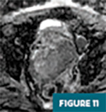
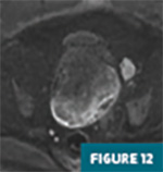
Pelvic MRI T2-weighted sagittal fast spin echo (FSE) (Figures 1-4) and T2-weighted axial FSE (Figures 5-8) images revealed an inverted uterus secondary to an 8.7 cm x 8.4 cm solid, heterogenous, and hyperintense mass, centered in the upper and mid aspects of the uterus with associated displacement craniocaudally.

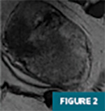
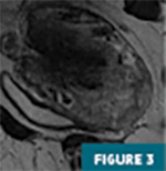
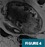
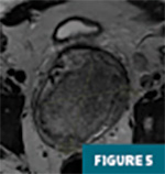
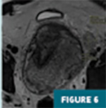
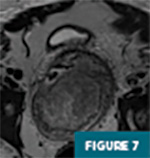
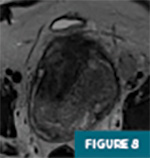
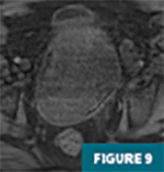
This mass demonstrated significant postcontrast enhancement. T1-weighted noncontrast axial FSE (Figure 9) and T1-weighted postcontrast axial FSE (Figure 10) images are provided for reference. This uterine mass also demonstrated restricted diffusion based on a decrease in signal on apparent diffusion coefficient (Figure 11) and hyperintense signal on diffusion- weighted imaging (Figure 12). A morphologically abnormal left external iliac lymph node measuring 2.4 cm in the short axis was noted (Figures 6 and 8), which was concerning for metastatic disease.

The histopathology of the mass revealed a partially necrotic, undifferentiated carcinoma with extensive myometrial invasion and focal lymphovascular invasion. Final immunohistochemical staining was consistent with a SMARCA4-deficient uterine sarcoma.
Diagnosis
Nonpuerperal prolapsed uterine inversion secondary to a uterine sarcoma
Discussion
Uterine inversion is a rare condition that occurs when the fundus of the uterus collapses into the uterine cavity.1 Uterine inversion can be a potentially life-threatening emergency.2 Without an accurate diagnosis and timely management of acute cases of uterine inversions, this condition can lead to persistent blood loss, hemorrhagic shock, and tissue necrosis.1 The incidence of puerperal uterine inversion is estimated to be 1/30,000 deliveries.3 Nonpuerperal uterine inversion is a rarer condition occurring in approximately 17% of all uterine inversions.4 There were only 190 cases reported in the literature from 1940 to 2020, and the mean age of the patient population was 47.90 ± 17.2 years.5 Nonpuerperal uterine inversion can be caused idiopathically but also can occur as a sequela of a benign or malignant tumor within the uterine body.3
Nonpuerperal uterine inversion secondary to a uterine sarcoma is even more rare—5.6% of total reported cases of nonpuerperal uterine inversions.6 Most of the published literature on nonpuerperal uterine inversion is in the form of case reports. There are many proposed theories on how these occur. One proposed theory associated with sarcomas is that the uterine wall has been weakened by the mass, causing the uterus to prolapse into its cavity.6
There are four degrees of uterine inversions: incomplete (1st degree), complete (2nd degree), prolapsed (3rd degree), and total (4th degree).7 An incomplete uterine inversion occurs when the fundus folds on itself within the uterine cavity. A complete uterine inversion occurs when the fundus protrudes through the cervical os. A prolapsed uterine inversion occurs when the fundus is present within the vaginal introitus. A total uterine inversion occurs when both the uterus and the vagina are inverted. Uterine inversion can be further classified into two types: puerperal or obstetric and nonpuerperal or gynecological.8
Uterine sarcoma is a rare, aggressive, and malignant tumor with a poor prognosis. Uterine sarcomas are often diagnosed in postmenopausal women. The pathophysiology of uterine sarcomas remains unclear.9 Due to their rarity, similar clinical symptoms, and lack of specific diagnostic techniques, uterine sarcomas can occasionally be misdiagnosed as uterine fibroids, leading to delayed diagnosis in the advanced stages,9,10 as seen in this case. Therefore, differentiating between the two is vital.
This case depicts a nonpuerperal 3rd degree or prolapsed uterine inversion. The diagnosis can often be made clinically with a bimanual examination and confirmed with ultrasound.6 In this patient, bimanual examination could not be performed due to the patient’s bodily habitus, and ultrasound did not confirm uterine inversion secondary to a poor acoustic window. Since uterine sarcomas are rare, especially in the younger population, these could be overlooked and excluded in a radiologist’s differential diagnosis when utilizing ultrasound. Therefore, it is noteworthy that radiologists should consider a potential uterine sarcoma in their differential diagnosis because complications can be dangerous for the patient.
In our study, the patient suffered from PCOS; heavy, persistent vaginal bleeding resulting in hemorrhagic shock; endometrial thickening; and uterine sarcoma with tissue necrosis and metastasis to the left external iliac lymph node. This case depicts a unique and unusual presentation of a young woman with a rare nonpuerperal uterine inversion secondary to a necrotizing uterine sarcoma, initially misdiagnosed as a fibroid uterus. Initial physical examination findings were limited, due to the patient’s bodily habitus. Ultrasound findings revealed a highly limited examination but reported a fibroid uterus with either one large fibroid or multiple fibroids. MRI revealed a uterine inversion secondary to a morphologically malignant uterine mass. Final pathology and immunohistochemical staining were consistent with a malignant uterine sarcoma.
This particular case emphasizes important factors to consider. Prompt diagnosis of uterine inversion is vital, as early detection and intervention can reduce the complications. If the results from a pelvic ultrasound are inconclusive or limited, a repeat ultrasound or MRI imaging should be recommended to confirm the diagnosis of uterine inversion. Optimal treatment for uterine inversion secondary to uterine sarcoma is surgical management with removal of the mass. In this specific case, due to the size of the mass, persistent vaginal bleeding, and the patient’s high BMI, conservative management was initiated, which included bariatric surgery, chemotherapy, and radiation.
Adrian Torres, BS, is a third-year medical student at Florida State University College of Medicine in Tallahassee.
Sophia Mourad, BS, is a third-year medical student at Florida State University College of Medicine.
Andrew Dakkak, MD, is a diagnostic radiology resident at AdventHealth Orlando.
Nicholas Feranec, MD, is a body imaging diagnostic radiologist at AdventHealth Orlando.
References
1. Thakur M, Thakur A. Uterine Inversion. In: StatPearls [Internet]. Treasure Island, FL: StatPearls Publishing; 2022.
2. Wendel MP, Shnaekel KL, Magann EF. Uterine inversion: a review of a life-threatening obstetrical emergency. Obstet Gynecol Surv 2018;73(7):411-417.
3. Das P. Inversion of the uterus. BJOG. 1940;47(5):525-548.
4. Leconte I, Thierry C, Bongiorno A, Luyckx M, Fellah L. Non-puerperal uterine inversion. J Belg Soc Radiol. 2016;100(1):47.
5. Liu H, Bi Z, Hu Q, Liu S, Dong Z, Wang J. Non-puerperal uterine inversion with endometrial polyps in an 11-year-old girl: a case report. J Pediatr Adolesc Gynecol. 2022;35(2):188-191.
6. Herath RP, Patabendige M, Rashid M, Wijesinghe PS. Nonpuerperal uterine inversion: what the gynaecologists need to know? Obstet Gynecol Int. 2020;2020:8625186.
7. Skinner GN, Louden KA. Non-puerperal uterine inversion associated with an atypical leiomyoma. Aust N Z J Obstet Gynaecol. 2001;41(1):100-101.
8. Jones HW Jr. Non-puerperal inversion of the uterus. Am J Surg. 1951;81(5):492-495.
9. Kim JH, Kim HJ, Kim SH, et al. Sonographic and clinical characteristics of uterine sarcoma initially misdiagnosed as uterine fibroid in women in the late reproductive age. J Menopausal Med. 2019;25(3):164-171.
10. Bužinskienė D, Mikėnas S, Drąsutienė G, Mongirdas M. Uterine sarcoma: a clinical case and a literature review. Acta Med Litu. 2018;25(4):206-218.